

Managing Editor
@ShahidNShah
@ShahidNShah
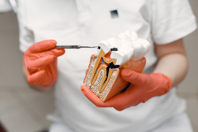
Few dental procedures carry as much undeserved anxiety as root canal therapy. That reputation largely belongs to a different era — one before digital imaging, precision rotary instruments, and refined anesthetic techniques transformed what was once a grueling multi-session ordeal into a procedure most patients describe as no worse than a filling. Yet the old narrative persists, and it sends people delaying care until a salvageable tooth becomes an extractable one.
The clinical stakes are real. When pulp tissue inside a tooth becomes irreversibly inflamed or infected — from deep decay, a crack, or repeated dental work — bacteria can spread into the surrounding bone if left untreated. Root canal therapy stops that progression while preserving your natural tooth, which matters more than most patients realize. Natural teeth distribute biting forces in ways that implants and bridges approximate but don’t fully replicate. Understanding what modern treatment actually involves — mechanically, experientially, and in terms of outcomes — makes it far easier to approach the procedure with accurate expectations rather than inherited fear.
Root canal therapy’s reputation was shaped by decades of limited anesthesia, hand instrumentation, and multi-visit protocols that gave infection and discomfort time to compound between appointments. Contemporary endodontics looks fundamentally different, and those differences have measurable consequences for both the procedure itself and long-term tooth survival.
On the diagnostic side, cone beam computed tomography (CBCT) has changed what clinicians can see before they ever pick up an instrument. Traditional periapical radiographs show canals in two dimensions, which means curved roots, extra canals, and subtle fractures frequently go undetected. CBCT produces three-dimensional volumetric images that reveal canal anatomy with a precision that substantially reduces surprises mid-procedure. This matters most for molars, where a missed fourth canal is one of the more common reasons a tooth fails to heal.
Instrumentation has advanced just as dramatically. Nickel-titanium (NiTi) rotary files replaced rigid stainless steel hand files as the standard for canal shaping. NiTi alloys flex with the natural curves of a root rather than forcing straight-line access, which reduces both procedural errors and the physical demands placed on the tooth itself. Combined with electronic apex locators — devices that determine working length (how deep to clean) with high accuracy — these instruments allow clinicians to clean canals thoroughly without risking perforation through the root tip.
At the sealing stage, bioceramic sealers have emerged as a significant upgrade over earlier epoxy-based materials. These calcium silicate–based compounds are biocompatible, dimensionally stable, and bond chemically to both the canal wall and the gutta-percha core material, creating a seal that resists bacterial microleakage more effectively than older formulations.
The practical result of these advances is that single-visit root canal therapy is now viable for the majority of uncomplicated cases, reducing total chair time and eliminating the treatment gap between appointments where reinfection can occur.
Root canal therapy follows a logical sequence: diagnose and prepare the tooth, clean and shape the canal system, then seal it permanently. Each phase has distinct clinical steps that patients rarely learn about in advance — which is part of why the experience feels opaque. Breaking it down makes the visit far less abstract.
Before treatment begins, the clinician establishes a clear indication for therapy. Irreversible pulpitis (pulp inflammation that won’t resolve on its own) and pulp necrosis (dead pulp tissue, often with periapical infection visible on imaging) are the two primary clinical triggers. Diagnosis involves a combination of percussion testing, thermal pulp testing, and radiographic review — and increasingly, CBCT when anatomy is complex or a previous restoration obscures the picture.
Once treatment is confirmed, rubber dam isolation is placed over the tooth. This step gets little attention in patient-facing guides, but it’s clinically essential: the rubber dam prevents irrigating solutions and small instruments from entering the airway, keeps saliva (and its bacterial load) out of the canal, and gives the clinician a clean working field. It’s also the moment when many patients relax, because the dam’s frame creates a physical boundary that makes the procedure feel contained.
Working length determination follows access preparation. The clinician uses an electronic apex locator — a small device that sends a mild electrical signal through a file placed in the canal — to establish precisely how deep cleaning should extend. This replaced the older method of repeated radiographic estimation and has made the process meaningfully more accurate.
With working length established, NiTi rotary instrumentation progressively enlarges and shapes the canal space. These files work in a continuous or reciprocating rotation driven by a handpiece, moving efficiently through the canal while conforming to its natural curvature. Irrigation runs continuously throughout this phase, most commonly with sodium hypochlorite solution, which dissolves organic debris, kills bacteria, and penetrates areas that instruments can’t reach mechanically. A final rinse with EDTA solution removes the smear layer left by instrumentation, exposing dentinal tubules for better sealer penetration.
Obturation — sealing the cleaned canal — follows immediately in single-visit cases, or after a temporary antimicrobial dressing in multi-visit protocols. The standard technique uses gutta-percha, a rubber-like material that is thermoplasticized and compressed to fill the canal space, combined with a sealer to close any voids. The choice between single-visit and multi-visit treatment isn’t arbitrary: teeth with active, draining infection may benefit from an interim calcium hydroxide dressing to further reduce bacterial load before final obturation. For most patients with vital or recently necrotic pulp and no acute abscess, single-visit therapy is entirely appropriate.

Knowing the mechanical sequence of a root canal helps intellectually — but most people want to know what they’ll actually feel at each stage. That’s a separate and equally important question.
Modern local anesthesia protocols have made root canal therapy reliably comfortable for the vast majority of patients. The standard approach uses inferior alveolar nerve blocks for mandibular (lower) teeth and infiltration injections for maxillary (upper) teeth, delivering profound pulpal anesthesia within minutes. Most patients feel pressure and movement during the procedure, not pain — and that distinction is worth holding onto.
The challenging cases are teeth in acute pulpitis, where tissue inflammation lowers the pH around the nerve and reduces local anesthetic efficacy. Clinicians address this with supplemental techniques: intraosseous injections deliver anesthetic directly into the bone adjacent to the tooth’s apex, producing near-instant profound numbness even in inflamed tissue. Intrapulpal injection is another option once access is established. These supplemental approaches have largely eliminated the scenario where a patient feels everything despite multiple standard injections.
For patients with significant dental anxiety, sedation options range from nitrous oxide (inhaled, wears off within minutes of stopping, allows the patient to drive afterward) to oral conscious sedation administered before the appointment. Some specialty practices offer IV sedation. In-chair comfort measures — offering headphones, explaining each step before beginning it, taking breaks on request — meaningfully reduce perceived discomfort independent of pharmacology. If you’re concerned about anxiety management, consulting with an endodontist in New City before your appointment to discuss sedation options is a reasonable first step.
Postoperative soreness is normal and expected, primarily because the procedure involves instruments working near the periapical tissues at the root tip. Most patients experience mild to moderate tenderness to biting pressure for two to three days, with full resolution typically within a week for uncomplicated cases.
Evidence-based analgesic protocols generally favor ibuprofen as first-line management, used around the clock at appropriate doses for the first 24 to 48 hours rather than waiting for pain to peak before medicating. Combining ibuprofen with acetaminophen provides additive relief through different mechanisms, and research published in the *Journal of the American Dental Association* supports this combination as more effective than either drug alone for postoperative endodontic pain.
Antibiotics are indicated only when infection has spread systemically (fever, swelling, lymphadenopathy) — not for routine prophylaxis. Using them otherwise contributes to resistance without benefit. Red flags that warrant prompt follow-up include swelling that spreads into the neck or floor of the mouth, fever above 101°F, or pain that intensifies rather than gradually diminishes after the first 48 hours.
A 2016 systematic review published in the *Journal of Endodontics* reported pooled success rates for initial root canal treatment in the range of 86–98%, with variation depending on preoperative diagnosis, tooth type, and whether periapical pathology was present at the time of treatment. Those numbers hold up well in long-term follow-up studies, particularly when the tooth receives an adequate coronal restoration — typically a crown — within a reasonable time after the root canal is completed. Leaving a treated tooth without proper coronal coverage is one of the clearest predictors of failure.
Complications do occur and are worth understanding honestly. Instrument separation — a file tip fracturing inside the canal — happens in a small percentage of cases, more often in calcified or severely curved canals. Depending on location, separated fragments can sometimes be retrieved or bypassed; in other cases they’re left in place with good clinical outcomes. Perforation through the root wall is rare with modern apex locators but remains a recognized risk. Persistent periapical infection that doesn’t resolve despite technically adequate treatment indicates a need for endodontic retreatment (non-surgical revision) or, in some cases, apicoectomy — a minor surgical procedure that removes the root tip and seals it from the outside.
The extraction-versus-treatment question deserves an honest framing. Dental implants have strong long-term data and can replace an extracted tooth with good functional outcomes — but they require surgery, osseointegration time, and typically greater cost than root canal therapy plus a crown. Preserving a natural tooth, when anatomy and prognosis support it, remains the first clinical preference across major endodontic guidelines.
The most useful thing to ask your clinician before treatment isn’t “will it hurt?” but rather “what does this tooth’s anatomy show on imaging, and what’s the prognosis for long-term retention?” That question frames the conversation around the information that actually drives the decision.

Most parents track their child’s height on a doorframe, but almost no one tracks jaw growth — and that gap has real consequences. The face and jaws develop more rapidly between birth and age …
Posted Sep 13, 2026 Dental Care Dentistry
Connecting innovation decision makers to authoritative information, institutions, people and insights.

Medigy accurately delivers healthcare and technology information, news and insight from around the world.
Medigy surfaces the world's best crowdsourced health tech offerings with social interactions and peer reviews.
© 2026 Netspective Foundation, Inc. All Rights Reserved.
Built on Oct 7, 2026 at 6:05pm