

Managing Editor
@ShahidNShah
@ShahidNShah
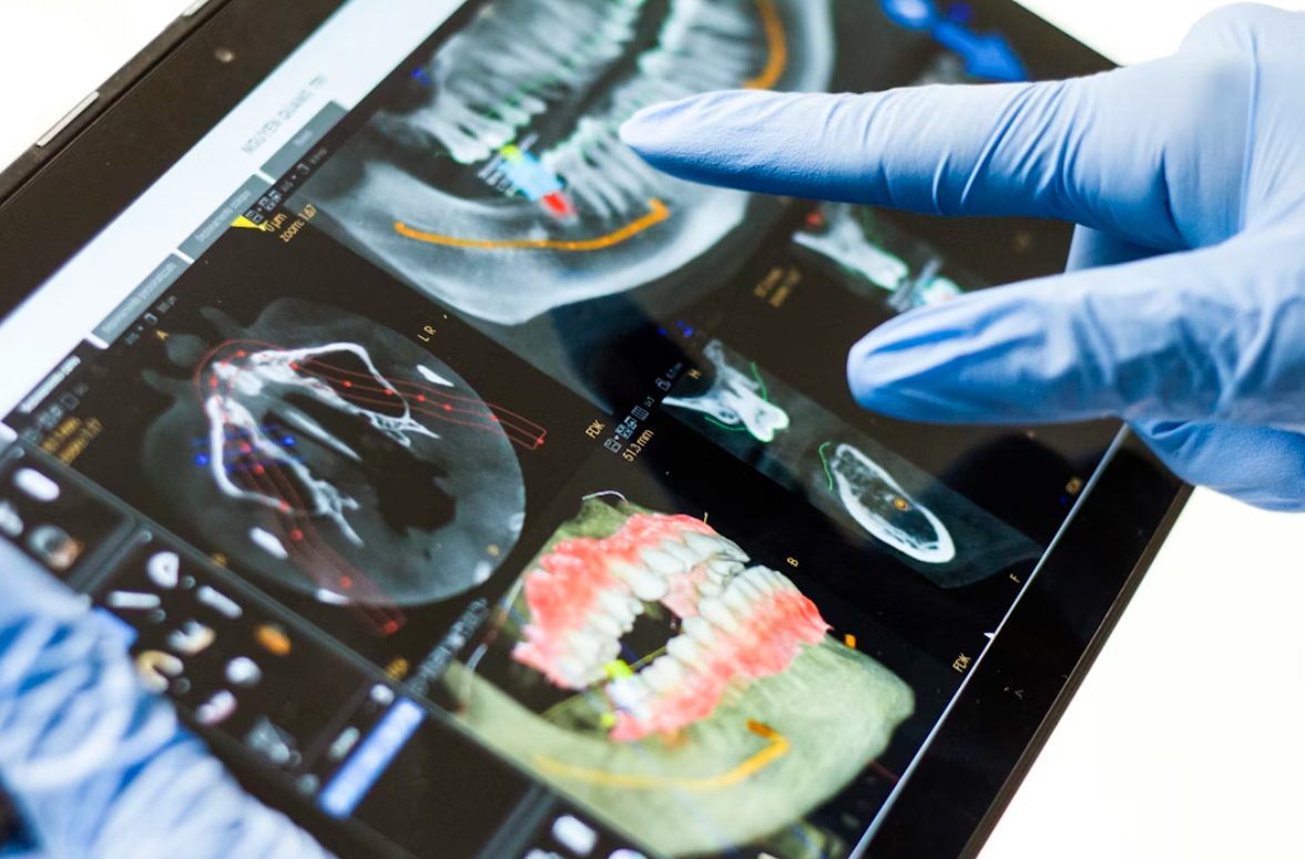
A flat X-ray tells you a tooth is missing and roughly where the bone sits. What it can’t tell you is whether that bone is thick enough at the exact point an implant needs to go or whether a nerve takes an unexpected path through the area. Those are the questions that determine whether surgery goes smoothly, and they’re invisible in two dimensions.
That gap is the reason 3D scanning became standard in implant planning rather than staying a specialty add-on. Practices in Charleston increasingly scan before committing to any surgical plan for exactly this reason.
Here’s what the imaging actually reveals while there’s still time to change course.
Two-dimensional radiography has known structural limitations. An evidence-based review published through the NIH’s National Center for Biotechnology Information concluded there is significant evidence supporting cone beam CT in preoperative assessment for defining pertinent anatomy, alveolar bone volume, and precise implant size selection, while noting panoramic imaging is unreliable unless meticulous positioning precautions are taken.
The specific failures are magnification error, distortion, and superimposition of structures on top of one another. A panoramic image compresses a curved three-dimensional jaw onto a flat plane, which means bone that appears adequate may be adequate in only one direction.
The inferior alveolar nerve running through the lower jaw is the structure surgeons most need to avoid, and its path is less predictable than standard anatomy implies. Diagrams in a textbook show a relatively consistent, single-canal route, but real anatomy frequently deviates from that idealized picture in ways that only become apparent through direct imaging.
That difference is striking and clinically significant. A branching nerve invisible on a flat film sits directly in the path of a planned implant in some patients, and nerve injury during implant placement can produce lasting numbness of the lip and chin. Knowing the anatomy beforehand is the entire point, since a plan built without this information risks discovering the variation only after the damage has already occurred.
Bone varies in density between individuals and between regions of the same jaw, and that variation affects how an implant is placed and how long it needs before loading.
This assessment is one reason patients considering dental implants in Charleston may receive a treatment timeline tailored to their individual needs rather than a standard schedule. By using 3D imaging to evaluate the available bone and surrounding anatomy, practices such as Charleston Center for Cosmetic and Restorative Dentistry can develop a more personalized treatment plan based on each patient’s specific circumstances.
Upper posterior implants sit close to the maxillary sinus, and the amount of bone between the ridge and the sinus floor determines whether a straightforward placement is possible or whether a sinus lift is required first. This measurement is essentially invisible on a standard exam, which is exactly why imaging becomes necessary before any upper posterior implant is planned.
Scanning also reveals sinus health, since existing inflammation or pathology needs addressing before any procedure that involves the sinus membrane. Discovering that during planning means a referral and a delay. Discovering it during surgery means a considerably worse day for everyone.
Implants need bone on all sides, and the dimension that most often falls short is width rather than height. A ridge can be perfectly tall and too thin to house an implant without the surface perforating.
Two-dimensional imaging shows height and says nothing about width, which is why patients occasionally arrive for surgery only to have the plan change mid-procedure. A scan measures buccal and lingual bone thickness directly, meaning any need for grafting is identified during planning rather than discovered with the site already open.
The scan doesn’t just inform the plan; it can produce a physical guide.
The strongest case for scanning remains complex anatomy rather than every routine case.
The genuine value of scanning is that it moves surprises from the operating chair to the planning stage, where they’re cheap to address. Insufficient bone width, an unexpected nerve branch, a sinus that needs attention, and all of these are manageable problems when identified in advance and considerably worse when discovered mid-procedure. If you’re being offered implants without any three-dimensional assessment, it’s reasonable to ask why, particularly for lower posterior sites where nerve proximity is the concern.
It’s equally reasonable for a dentist to answer that your case is straightforward enough not to require it, since additional imaging means additional radiation exposure and shouldn’t be automatic. What matters is that the decision was considered rather than skipped.

Two patients sit in the same waiting room. Same age, same insurance, same twice-yearly cleaning schedule. One will develop three cavities over the next four years, and the other won’t develop …
Posted Aug 17, 2026 Dental Care Dentistry
Connecting innovation decision makers to authoritative information, institutions, people and insights.

Medigy accurately delivers healthcare and technology information, news and insight from around the world.
Medigy surfaces the world's best crowdsourced health tech offerings with social interactions and peer reviews.
© 2026 Netspective Foundation, Inc. All Rights Reserved.
Built on Oct 18, 2026 at 6:05pm