

Managing Editor
@ShahidNShah
@ShahidNShah
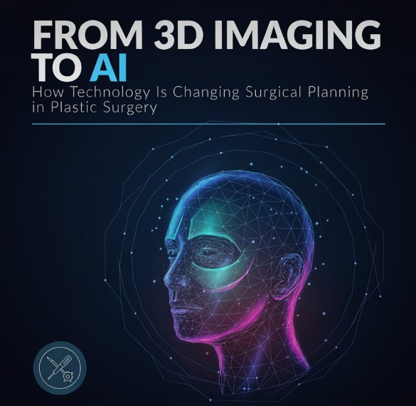
Plastic surgery has always depended heavily on visual assessment. Surgeons evaluate proportions, symmetry, tissue quality, anatomical relationships and how changes to one area may affect the appearance of another.
Historically, much of that planning has relied on physical examination, standard photography, measurements and the surgeon’s experience.
Those fundamentals remain important, but the tools available before a patient enters the operating room are becoming considerably more sophisticated.
Three-dimensional imaging can create detailed representations of a patient’s anatomy. Digital simulations can help surgeons and patients explore potential changes before a procedure. Artificial intelligence is being investigated for applications ranging from automated anatomical measurements to risk prediction and postoperative assessment.
The result is not autonomous surgery or a replacement for clinical judgment. Instead, technology is increasingly providing surgeons with additional information that can make preoperative planning more quantitative, visual and individualized.
Standardized clinical photography remains an important part of plastic surgery, but photographs inherently reduce three-dimensional anatomy to a flat image.
That limitation matters when evaluating structures defined by volume and projection. Breast asymmetry, facial contours and changes in the abdominal or truncal surface, for example, may be difficult to quantify precisely using photographs alone.
Three-dimensional surface imaging addresses some of these limitations by capturing the geometry of the body or face from multiple angles and producing a digital model. Depending on the system and procedure, surgeons can use these models to evaluate differences in volume, contour, projection and symmetry.
The technology has found applications in both reconstructive and aesthetic surgery. Three-dimensional imaging can assist with facial analysis, breast surgery and assessment of postoperative changes, while CT- and MRI-derived 3D reconstructions can provide additional anatomical information in complex reconstructive cases.
The value is not simply creating a more impressive image for the patient. Quantifiable measurements can supplement the surgeon’s physical examination and provide a reproducible baseline against which subsequent changes can be assessed.
One of the most visible applications of 3D technology is surgical simulation.
In procedures such as breast augmentation or rhinoplasty, software can manipulate a patient’s three-dimensional model to demonstrate how different changes could affect overall proportions. These simulations can make conversations about size, projection and contour easier for patients to understand than descriptions based entirely on measurements or photographs.
But simulation introduces an important distinction: visualization is not prediction.
A computer-generated image cannot fully account for variables such as tissue elasticity, wound healing, scar formation, postoperative swelling or individual biological responses to surgery. A simulated result should therefore be understood as a communication and planning tool rather than a guarantee of the surgical outcome.
That distinction becomes increasingly important as simulations become more realistic.
“Technology can help us measure anatomy and communicate a surgical plan much more clearly, but it doesn’t change the biological variability we deal with in surgery,” says plastic surgeon Dr. Waqqas Jalil. “Two patients can have similar measurements and still have differences in skin quality, tissue characteristics and healing. The imaging has to be interpreted in the context of the actual patient.”
Artificial intelligence potentially takes digital surgical planning a step further.
Rather than simply creating a three-dimensional representation, machine-learning systems can analyze large quantities of imaging and clinical data to identify patterns that may be difficult to recognize consistently through manual assessment.
Researchers have investigated AI applications in facial landmark identification, image segmentation, aesthetic assessment, outcome prediction and surgical risk stratification. In reconstructive surgery, AI-assisted imaging is also being studied for applications such as vascular mapping and flap planning.
A 2025 systematic review published in Frontiers in Surgery evaluated 25 studies examining AI applications in plastic surgery. The researchers reported promising performance in preoperative planning, predictive modelling and postoperative evaluation, while also identifying significant limitations in the available evidence.
That combination of promise and uncertainty is important. AI systems may perform impressively on the datasets used to develop them without necessarily producing the same results across different patient populations or clinical environments.
Not every technological advance in surgical planning is focused on appearance.
Machine learning can potentially analyze clinical variables to estimate the probability of complications or identify patients with characteristics associated with particular outcomes. This creates another possible role for AI before surgery: supporting risk assessment.
Traditional surgical risk assessment already considers factors such as age, medical history, smoking, body mass index, medications and previous operations. Machine-learning models can theoretically examine relationships among a much larger number of variables simultaneously.
For plastic surgeons, such systems could eventually supplement clinical decision-making when determining whether a procedure is appropriate, whether several procedures should be combined or whether a patient’s risk profile warrants a different approach.
The key word, however, is supplement.
Clinical decisions involve factors that may not be represented adequately in a dataset. A probability generated by an algorithm is not equivalent to a comprehensive medical assessment, particularly when the system has not been validated in a population comparable to the patient being treated.
AI is only as useful as the information on which it has been trained.
That presents a particularly interesting problem in plastic surgery because human anatomy and aesthetic characteristics vary substantially across age groups, sexes, ethnic backgrounds and body types.
If an image-analysis model is trained predominantly on a narrow population, its measurements or recommendations may be less reliable when applied to patients who are poorly represented in that dataset.
Recent reviews of AI in plastic surgery have repeatedly identified dataset diversity, external validation and algorithmic bias as barriers to widespread clinical implementation.
There is also a more philosophical issue in aesthetic surgery. Algorithms trained to evaluate attractiveness, symmetry or desirable postoperative outcomes inevitably encode assumptions about what constitutes an aesthetically preferable result.
Plastic surgery is not simply an exercise in maximizing mathematical symmetry. Patient preferences, cultural differences, anatomical feasibility and preservation of individual characteristics all matter.
The most realistic future for technology in plastic surgery is therefore probably not one in which an algorithm determines what operation a patient should have.
Instead, surgeons are gaining a larger collection of digital tools.
Three-dimensional imaging can provide additional anatomical measurements. Simulation can improve communication. AI may help identify patterns in imaging or clinical data. Augmented-reality systems are being investigated as a way to bring elements of preoperative planning into the operating room itself.
Research is moving quickly, but clinical adoption should move according to evidence rather than novelty.
A recent systematic review of AI, augmented reality and robotics in aesthetic practice found considerable potential for technologies including image analysis, volumetric planning and intraoperative navigation, but characterized the overall methodological quality of the available evidence as low to moderate.
That gap between technological capability and clinical validation will be one of the central challenges as these systems become more accessible.
Plastic surgery will remain a field in which technical skill, anatomical knowledge and clinical judgment are difficult to separate from the quality of the result.
What is changing is the amount of information available to the surgeon before making those decisions.
The transition from photographs and manual measurements to 3D anatomical models, quantitative image analysis and increasingly sophisticated AI systems gives surgeons new ways to examine anatomy and communicate treatment plans.
The technology is unlikely to make plastic surgery completely predictable. Human anatomy and healing are far too variable for that.
Its more practical contribution may be making surgical planning better informed: giving surgeons additional ways to measure what they see, evaluate potential approaches and explain those decisions to patients before entering the operating room.


In the past, visiting the doctor used to mean one thing. You walked in, waited your turn, and walked out with a prescription. That is no longer how most people experience healthcare. Today, a single …
Posted Aug 29, 2026 Patient Experience
Connecting innovation decision makers to authoritative information, institutions, people and insights.

Medigy accurately delivers healthcare and technology information, news and insight from around the world.
Medigy surfaces the world's best crowdsourced health tech offerings with social interactions and peer reviews.
© 2026 Netspective Foundation, Inc. All Rights Reserved.
Built on Oct 14, 2026 at 6:06pm